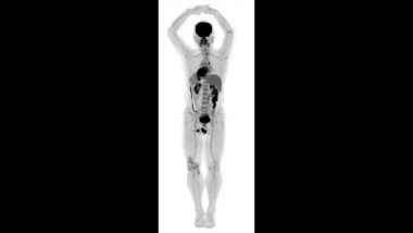
Los Angeles, November 20: The world's first medical imaging scanner that can capture a 3D picture of the whole human body at once in as little as 20-30 seconds, has produced its first scans, scientists said Monday. Developed by scientists from the University of California - Davis in the US, EXPLORER is a combined positron emission tomography (PET) and X-ray computed tomography (CT) scanner that can image the entire body at the same time.
Since the machine captures radiation far more efficiently than other scanners, EXPLORER can produce an image in as little as one second and, over time, produce movies that can track specially tagged drugs as they move around the entire body.
The technology will have countless applications, from improving diagnostics to tracking disease progression to researching new drug therapies, researchers said. Social Media Addicted? Maharashtra’s First Internet De-Addiction Centre Set Up in Pune.
EXPLORER will have a profound impact on clinical research and patient care because it produces higher-quality diagnostic PET scans than have ever been possible, they said.
It also scans up to 40 times faster than current PET scans and can produce a diagnostic scan of the whole body in as little as 20-30 seconds, according to the researchers.
EXPLORER can scan with a radiation dose up to 40 times less than a current PET scan, they said. The scanner has been developed in partnership with Shanghai-based United Imaging Healthcare (UIH), which will eventually manufacture the devices for the broader healthcare market. World’s First Total-Body Scanner Reveals Pictures of Full Human Body Scan.
"While I had imagined what the images would look like for years, nothing prepared me for the incredible detail we could see on that first scan," said Simon Cherry, a professor at UC Davis.
"While there is still a lot of careful analysis to do, I think we already know that EXPLORER is delivering roughly what we had promised," Cherry said. The first images from scans of humans using the new device will be shown at the upcoming Radiological Society of North America meeting, which starts on November 24 in Chicago.
"The level of detail was astonishing, especially once we got the reconstruction method a bit more optimized," said Ramsey Badawi from UC Davis. "We could see features that you just don't see on regular PET scans. And the dynamic sequence showing the radiotracer moving around the body in three dimensions over time was, frankly, mind-blowing," Badawi said. "There is no other device that can obtain data like this in humans, so this is truly novel," he said.


 Quickly


