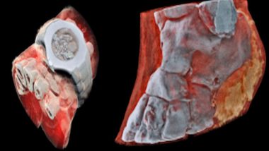
Gone are the days when a high-contrast, black-and-white image of your bones was used for detecting fractures or breaks in your body. Recently there is a remarkable invention that has come in which the X-ray will unveil much more than just the bones inside you. Yes you heard it right now your X-rays will be 3D and unlike earlier, you will get full-colour images of your X-rays. The primary purpose that these images will serve is that doctors will now have a chance to diagnose the problem without cutting it open
The primitive method of imaging the insides of a patient only involved examining them with x-rays. The electromagnetic radiation has a shorter wavelength as compared to visible light, so it would pass soft tissues easily but failed to pass through harder elements like bones. It thus exhibits an image based on the intensity of the x-rays that make it through, further detecting fractures or breaks in your body on a film which is placed on the other side of your body.
A company based out in New Zealand named Mars Bioimaging has revealed an innovative type of medical imaging scanner which operates similarly but adopts technology produced for the Large Hadron Collider at CERN to generate considerably more detailed issues. The Medipix3 chip works equally with a sensor of a digital camera, but it identifies and calculates the particles are hitting each pixel when a shutter opens.
According to their official website MARS, scanners generate multi-energy images with high spatial resolution and low noise. Functional imaging simultaneously identifies and quantifies various components of soft tissues, bones, cartilage as well as exogenously administered contrast agents, nanoparticles and pharmaceuticals in a single scan.
They further describe their project by saying, The MARS small bore scanner enables customers to conduct experiments in a system that is directly translatable to human imaging. MBI works closely with customers to provide a wide range of spectral imaging solutions for applications ranging from cancer detection to development of novel contrast agents.
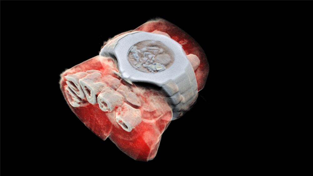
Here are a few pictures for reference:
Wrist:
Ankle left view:
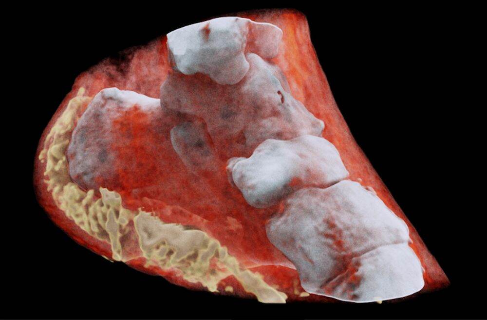
Ankle right view:
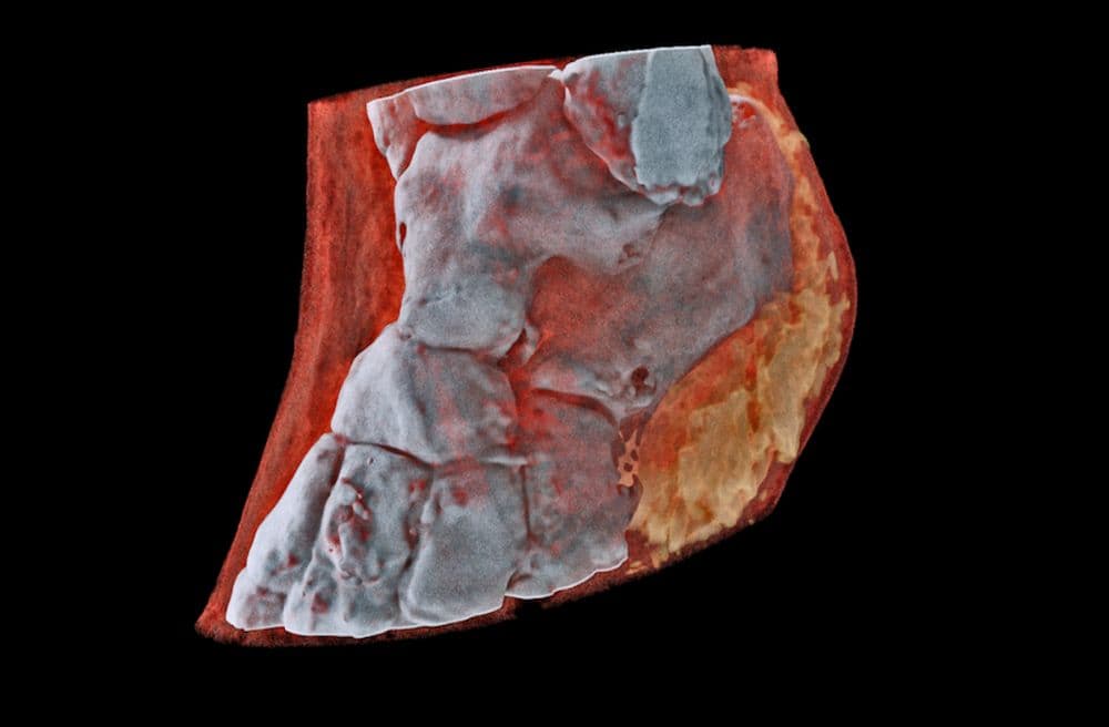
This surely seems to an amazing effort taken towards the diagnosis of various ailments and seems like the future of diagnosing cancer and various other health problems.
(The above story first appeared on LatestLY on Jul 14, 2018 12:31 AM IST. For more news and updates on politics, world, sports, entertainment and lifestyle, log on to our website latestly.com).


 Quickly


