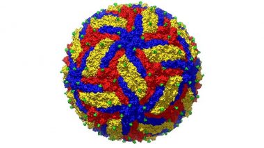
Is This How The Zika Virus Looks? High Resolution Image of The Pathogen Can Hold Clues To Its Prevention

Scientists have developed the most detailed image ever of the Zika virus. This could enable the development of a perfect vaccine.

Health & Wellness Team Latestly|
Is This How The Zika Virus Looks? High Resolution Image of The Pathogen Can Hold Clues To Its Prevention
Is this the best image of the Zika virus. Scientists think so. (Photo Credits: Sevvana et. al)

In a reassuring move, scientists have developed a high-resolution image of the Zika virus, throwing open possibilities of fighting it more effectively. After scientists developed a technique to manipulate mosquito sperms to prevent Zika, researchers biologists Madhumati Sevvana and her colleagues have mapped out the most detailed image of the Zika virus, complete with the glycoprotein interactions and surface properties.

The findings of the study were published in the journal Structure, where the scientists combined thousands of two-dimensional images of the virus to construct a three-dimensional version of the virus. The feat deserves applause because this is the first time a zoomed-in, detailed image of a flavivirus, which includes Zika, dengue, yellow fever virus, has been developed.
Mr World 2024 Winner Is Danny Mejia From Puerto Rico; India’s Gokul Ganesan Leaves His Mark by Securing Spot in Top 10 (Watch Video)
  • Sobhita Dhulipala's Traditional Looks Prove That She'll Look Gorgeous on Her D-day (View Pics)
  • Close
    Search

    Is This How The Zika Virus Looks? High Resolution Image of The Pathogen Can Hold Clues To Its Prevention

    Scientists have developed the most detailed image ever of the Zika virus. This could enable the development of a perfect vaccine.

    Health & Wellness Team Latestly|
    Is This How The Zika Virus Looks? High Resolution Image of The Pathogen Can Hold Clues To Its Prevention
    Is this the best image of the Zika virus. Scientists think so. (Photo Credits: Sevvana et. al)

    In a reassuring move, scientists have developed a high-resolution image of the Zika virus, throwing open possibilities of fighting it more effectively. After scientists developed a technique to manipulate mosquito sperms to prevent Zika, researchers biologists Madhumati Sevvana and her colleagues have mapped out the most detailed image of the Zika virus, complete with the glycoprotein interactions and surface properties.

    The findings of the study were published in the journal Structure, where the scientists combined thousands of two-dimensional images of the virus to construct a three-dimensional version of the virus. The feat deserves applause because this is the first time a zoomed-in, detailed image of a flavivirus, which includes Zika, dengue, yellow fever virus, has been developed.

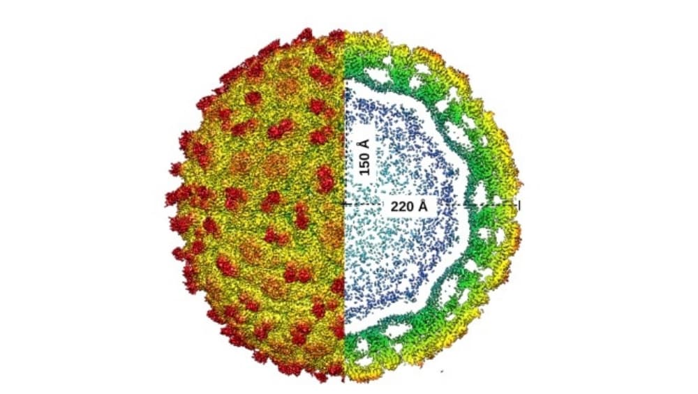
    Zika virus (Photo Credits: Sevvana et. al)

    They say the newly-developed structure has been developed using a cryo-electron microscopy, a method used for determining biomolecules in solution, to develop this highly-detailed image of the dangerous virus. By understanding the surface mechanism of the virus, the scientists will now be able to clearly see the drug-binding pockets on its surface, which could bind to the drug molecules. These pockets work as sockets, and the perfect vaccine will be the plug that could fit into these sockets, destroying the virus from within. It's just a matter of time before researchers develop the right vaccine.

    Additionally, the scientists also understood that each virus in the flavivirus family differs from each other in terms of structure. Each is different at specific regions of their surface called glycan loop. These sites determine which cell in the body the virus could infect and what symptoms they could cause. That’s why every flavivirus infection results in different symptoms. In Zika, it causes birth defects, and in dengue, it develops into a haemorrhagic fever.

    The discovery is a milestone in Zika virus treatment since the surface differences will enable scientists to find the perfect vaccine, changing the game of the treatment.

    (The above story first appeared on LatestLY on Jun 27, 2018 12:43 PM IST. For more news and updates on politics, world, sports, entertainment and lifestyle, log on to our website latestly.com).

    Asia's Chronic Pollution Has High Health Costs
    Science " onclick="shareOpen('https://twitter.com/intent/tweet?url=https%3A%2F%2Fwww.latestly.com%2Flifestyle%2Fhealth-wellness%2Fis-this-how-the-zika-virus-looks-high-resolution-image-of-the-pathogen-can-hold-clues-to-its-prevention-235492.html&text=Is+This+How+The+Zika+Virus+Looks%3F+High+Resolution+Image+of+The+Pathogen+Can+Hold+Clues+To+Its+Prevention&via=latestly', 650, 420);">
    Health & Wellness Team Latestly|
    Is This How The Zika Virus Looks? High Resolution Image of The Pathogen Can Hold Clues To Its Prevention
    Is this the best image of the Zika virus. Scientists think so. (Photo Credits: Sevvana et. al)

    In a reassuring move, scientists have developed a high-resolution image of the Zika virus, throwing open possibilities of fighting it more effectively. After scientists developed a technique to manipulate mosquito sperms to prevent Zika, researchers biologists Madhumati Sevvana and her colleagues have mapped out the most detailed image of the Zika virus, complete with the glycoprotein interactions and surface properties.

    The findings of the study were published in the journal Structure, where the scientists combined thousands of two-dimensional images of the virus to construct a three-dimensional version of the virus. The feat deserves applause because this is the first time a zoomed-in, detailed image of a flavivirus, which includes Zika, dengue, yellow fever virus, has been developed.

    Zika virus (Photo Credits: Sevvana et. al)

    They say the newly-developed structure has been developed using a cryo-electron microscopy, a method used for determining biomolecules in solution, to develop this highly-detailed image of the dangerous virus. By understanding the surface mechanism of the virus, the scientists will now be able to clearly see the drug-binding pockets on its surface, which could bind to the drug molecules. These pockets work as sockets, and the perfect vaccine will be the plug that could fit into these sockets, destroying the virus from within. It's just a matter of time before researchers develop the right vaccine.

    Additionally, the scientists also understood that each virus in the flavivirus family differs from each other in terms of structure. Each is different at specific regions of their surface called glycan loop. These sites determine which cell in the body the virus could infect and what symptoms they could cause. That’s why every flavivirus infection results in different symptoms. In Zika, it causes birth defects, and in dengue, it develops into a haemorrhagic fever.

    The discovery is a milestone in Zika virus treatment since the surface differences will enable scientists to find the perfect vaccine, changing the game of the treatment.

    (The above story first appeared on LatestLY on Jun 27, 2018 12:43 PM IST. For more news and updates on politics, world, sports, entertainment and lifestyle, log on to our website latestly.com).

    IPL Auction 2025 Live
    IPL Auction 2025 Live
    • INR
    • USD
    • EUR
    View all
    City Petrol Diesel
    New Delhi 96.72 89.62
    Kolkata 106.03 92.76
    Mumbai 106.31 94.27
    Chennai 102.74 94.33
    View all
    Currency Price Change